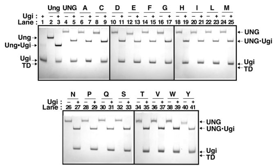
Fig. 3. Ability of UNG and R276X mutant proteins to bind Ugi.
Reaction mixtures (15 µl) containing 40 pmol of E. coli Ung (lanes 2 and 3), UNG (lanes 4 and 5), or R276X mutant protein (lanes 6–41), with or without (+ or −) Ugi (100 pmol) were incubated as described under “Experimental Procedures.” A control reaction containing Ugi (100 pmol) alone was similarly processed (lane 1). Samples were analyzed at 4 °C by non-denaturing 10% polyacrylamide gel electrophoresis, proteins were visualized with Coomassie Brilliant Blue G-250 stain, and the gel was imaged as described under “Experimental Procedures.” Arrows indicate the location of Ung, UNG, Ugi, Ung·Ugi, UNG·Ugi, and the tracking dye front (TD). Arg276 mutant protein lane assignments are indicated by single letter amino acid abbreviations.

