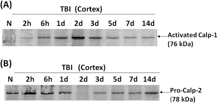
Figure 8. Examination of autolytic activation of calpain-1 and calpain-2 in rat cortex following TBI.
Western blot of naïve and cortex CCI time course samples similar to those in Figure 6. However, these blots were probed with polyclonal antibody specific to anti-activated calpain-1 new N-terminal (anti-NH2-LGRHENA) (A), or pro-calpain-2 N-terminal (anti-SHERAIK) (B). Results shown are representative of three separate experiments.

