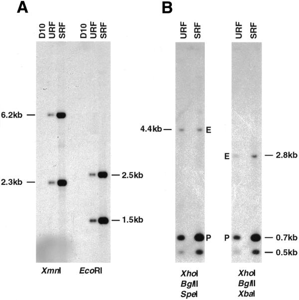
Figure 3.

Southern blot analysis of D10-ΔMSP1#2 gDNA following no (URF) or three cycles (SRF). (A) Parental D10, URF and SRF gDNA was digested with XmnI or EcoRI separated on 0.6% agarose gel and transferred to a nylon membrane. The filter was then hybridised with a TgDHFR-TS probe. (B) URF or SRF gDNA was digested with a combination of either XhoI, BglII, SpeI or XhoI, BglII, XbaI as indicated, electrophoresed on 0.6% agarose gel and transferred to nitrocellulose membrane. The filter was hybridised with the 900 bp MSP-1 probe and the signal quantitated by phosphorimager analysis. The larger band (4.4 or 2.8 kb) represents the single endogenous copy of the MSP-1 target sequence (E) while the 0.7 kb species corresponds to the XhoI–BglII fragment derived from the transfected plasmid (P).
