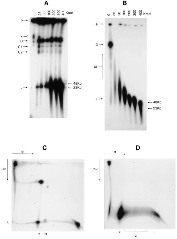
Figure 5.
PFGE analysis of the effect of ionising radiation from a 60Co source on transfected plasmids. The gels were electrophoresed, Southern blotted and hybridised to a pUC19 probe as described in the Materials and Methods. (A) URF and (B) SRF agarose blocks (P) exposed to 0–400 Krad γ-rays. (C) URF DNA 2D PFGE. An agarose block was first irradiated with 25 Krad and after PFGE, the resulting track was removed, exposed to 100 Krad and electrophoresed at 90° under the same conditions. (D) SRF DNA 2D PFGE. Conditions were the same as for (C), except that the initial agarose block was unirradiated before the first-dimension electrophoresis. Other labels are referred to in the text. Molecular weight markers are indicated to the right of (A) and (B).

