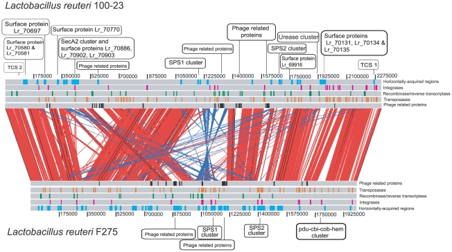
Figure 1. Pair-wise genomic comparisons between L. reuteri strains 100-23 and F275.
Linear genomic comparison of the chromosomes of 100-23 and F275 (using the sequence of JCM1112T). Both sequences are read left to right from the predicted origin of replication. Homologous regions within the two genomes identified by reciprocal BLASTN are indicated by red (same orientation) and blue (reverse orientation) bars. Putative horizontally acquired islands as identified by Alien_hunter (blue boxes), phage proteins (black boxes), transposases (orange boxes), and integrases (pink boxes) are indicated.

