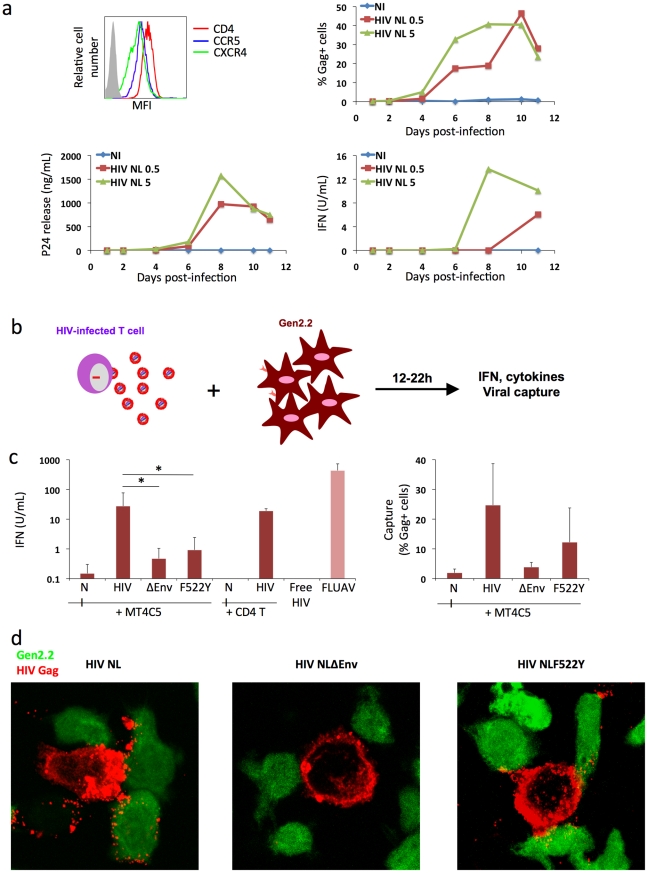
Figure 3. Recognition of HIV-infected cells by the Gen2.2 plasmacytoid-like cell line.
a. Gen2.2 cells express HIV receptors and are sensitive to HIV infection. Surface expression levels of CD4, CXCR4 and CCR5 are shown on the upper left panel. Gen2.2 cells were then exposed to HIV particles, NL4-3 strain, at two doses (0.5 and 5 ng/0.1 ml Gag p24/106 cells). Viral replication was assessed by following the appearance of Gag+ cells (upper right panel) and Gag p24 release in supernatants (lower left panel). IFN production was measured in the same supernatants (lower right panels). Data are representative of 3 independent experiments. b. Schematic representation of the coculture system. HIV-1 infected T cells are mixed with Gen2.2 cells and levels of IFN released in supernatants are measured 12–22 h later. c. Sensing of HIV-infected cells by Gen2.2 and role of viral envelope glycoproteins. MT4C5, infected with wild-type (HIV), Env-deleted (ΔEnv), or a non-fusogenic Env mutant (HIVF522Y), and with similar levels of Gag+ cells, were cocultivated with PBMCs and IFN was measured 22 h later (left panel). When stated, HIV-infected primary CD4+ cells were used as donors, instead of MT4C5 cells. Cell-free HIV virions (500 ng/ml p24) do not stimulate Gen2.2 cells. The efficiency of HIV capture by Gen2.2 was assessed by measuring the % of Gag+ cells by flow cytometry (right panel). Mean+sd of 10 independent experiments is shown, *p<0.05 (Kruskal-Wallis). d. Visualization of HIV capture by Gen2.2 cells. Gen2.2 cells were labeled with CFSE (green) and cocultured with MT4C5 infected with wild-type (HIV), ΔEnv and F522Y mutants. Cells were harvested after 1 h and stained for HIV Gag (red). Representative fields are shown.

