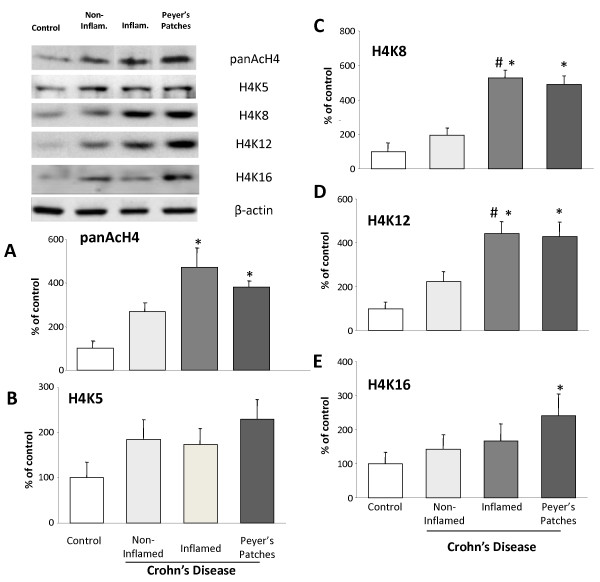
Figure 5.
Acetylation on histone 4 (H4) and H4 lysine residues in Crohn's disease. Columns represent the mean ± SEM of three independent experiments. Four biopsies were pooled to obtain sufficient protein for one experiment (50 μg of protein) (*p < 0.05 vs control). Pan acetylation on H4 in Crohn's disease (A). Acetylation on histone 4 (H4) specific lysine residues 5 (K5) (B), K8 (C), K12 (D), and 16 (E), in non-inflamed, inflamed tissue and Peyer's patches of Crohn's disease patients. Results were obtained by Western blotting. Columns represent the mean ± SEM of three independent experiments. (*p < 0.05 vs control, #p < 0.005 vs non-inflamed CD). Representative images of the bands obtained are illustrated.

