
Figure 5.
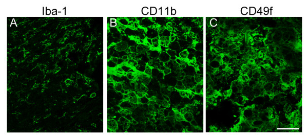
Immunofluorescence examination of Iba-1+ microglia, CD11b+ macrophages, CD49f+ macrophages/monocytes in the lesion center at day 14 post DSCI. The injured spinal cord tissue sections were collected at day 14 after SCI, and then subjected to immunofluorescence for Iba-1 (A), CD11b (B), or CD49f (C). Noted that the Iba1+ cells shown in A were located in the peritraumatic zone, and CD11b+/CD49+ cells in B and C were situated in the lesion center. Scale bar in A-F, 50 μm.