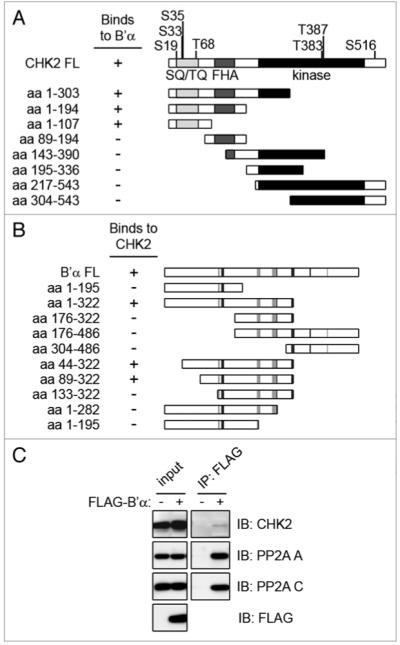
Figure 1.

CHK2 binds to the B’α regulatory subunit of pp2A. (A) the full length CHK2 protein is shown with phosphorylation sites and domains highlighted. the SQ/TQ domain is displayed in light gray, the FHA domain in dark gray, and the kinase domain in black. Using the yeast two-hybrid system, deletion mutants of CHK2 were co-transformed into yeast with full-length B’α. the minimal CHK2 binding region for B’α includes amino acid residues 1–107. (B) Full length B’α is shown with the residues responsible for binding to the pp2A A subunit in light gray and the C subunit in dark gray.23 Using the yeast two-hybrid system, deletion mutants of B’α were co-transformed with full-length CHK2. the minimal B’α binding region for CHK2 is comprised of amino acid residues 89–322. (C) Cell lysates of 293T cells expressing FLAG-B’α were immunoprecipitated (IP) using the FLAG antibody and immunoblotted (IB) for the indicated proteins. Cell lysates that were not expressing FLAG-B’α were used as a negative control.
