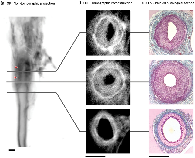
Figure 1. Identification of neointimal lesions in the ligation-injured mouse femoral artery.
In non-tomographic fluorescent emission images of a ligation-injured femoral artery (a) neointimal thickening is clearly visible (red arrowheads). Image has been inverted for clarity (darker regions reflect stronger emission). In tomographic reconstructions (b), all layers of the vessel wall can be identified. Reconstructions strongly resemble US trichrome-stained histological sections of the same vessels (c). Scale bars in (a–c) are 200 µm.

