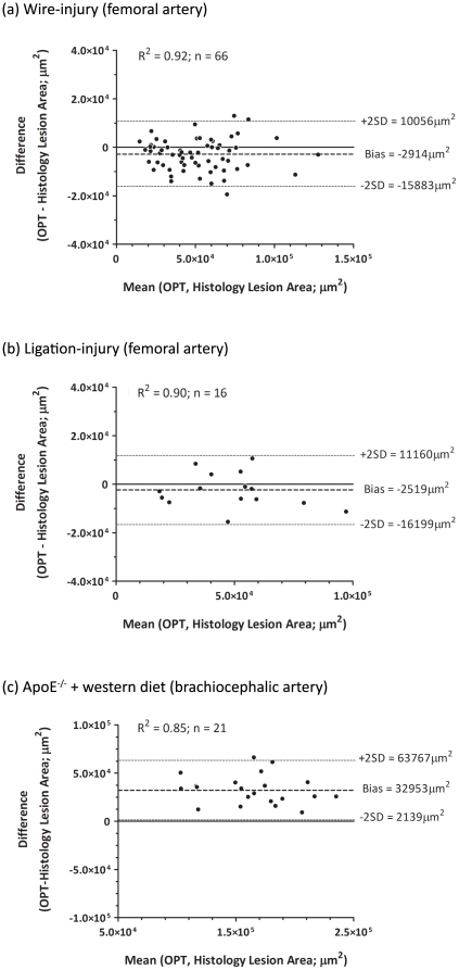
Figure 3. Comparison of planimetric measurements of lesion size between OPT reconstructions and histological sections.
Planimetric measurements of lesion size recorded from OPT and histology image sets correlate strongly for all lesion types; Bland-Altman analysis indicates that this is unbiased for wire- (a) and ligation-injured femoral arteries (b) and that OPT measurements have a consistent positive bias in atherosclerotic brachiocephalic arteries (c).

