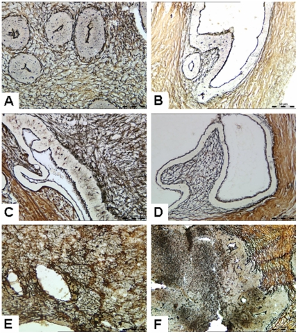
Figure 3. Reticular fiber staining of xenotransplanted human endometrial tissues.
(A) Human endometrial tissue before transplantation. (B) Control group. (C) 14d group. (D) 21d group. (E) 28d group. In the interstitial region, black fiber wire could be seen clearly, and the mesh structure remained intact before transplantation, or in the control group, or in the 14, 21 and 28 group. (F) 31d group. Black mesh fibers were broken in some parts of the interstitial region, fiber mesh structure disappeared, and no black filaments were observed in some areas. Figure is from serial thin sections. Original magnification: 400×.

