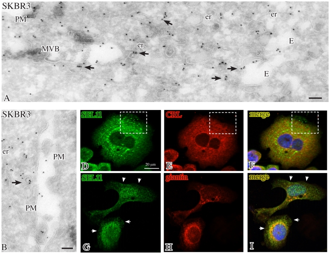
Figure 3. Localizations of SEL1L in untreated SKBr3 cells.
Cryoimmunogold electron microscopy of untreated SKBr3 cells labeled with 10 nm gold for N-terminal SEL1L (panels A, B) shows intense staining of vesicles (arrows) dispersed in the peripheral cytoplasm and sometimes associated with multivesicular bodies and endosomes. Immunofluorescence reveals that SEL1L (green) extensively colocalizes (yellow) with the endoplasmic reticulum marker calreticulin and, in a few dots, with the Golgi marker giantin (red), with the exception of discrete cytoplasmic foci (panels D–F, squares) and of the peripheral cytoplasm (panels G–I, arrows). Bars: 0.1 µm; E: endosomes; er: endoplasmic reticulum; MVB: multivesicular body; PM: plasma membrane.

