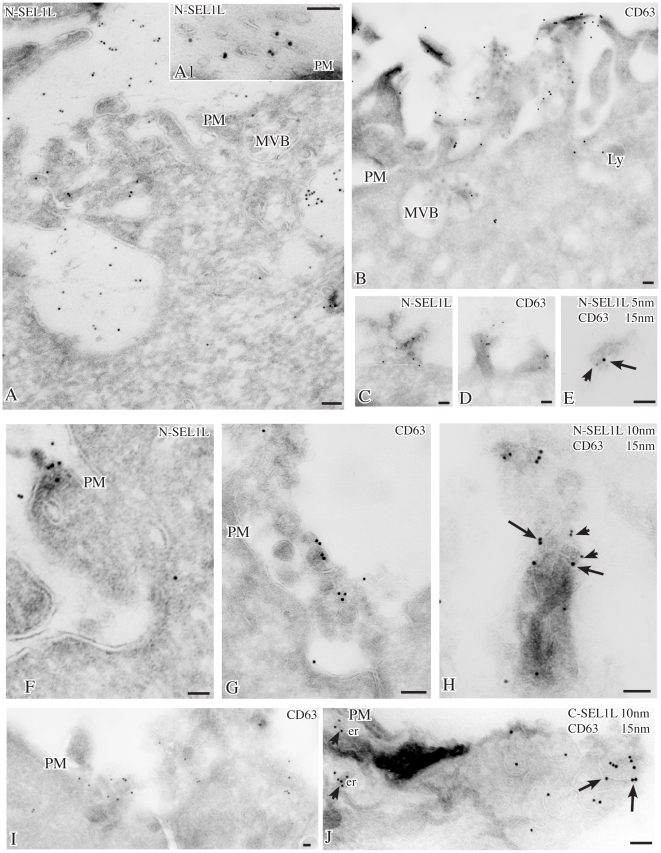
Figure 4. Localizations of SEL1L in DTT-treated SKBr3 cells.
After DTT treatment for 3 hrs, gold particles for N-terminal SEL1L (10 nm) are found by cryoimmunogold electron microscopy along the microvilli (panel A) and on membrane-bound vesicles in the extracellular space (panels A, A1) or emerging from the plasma membrane (panels C, F). Similarly, labeling for the tetraspan protein CD63 is detected in multivesicular bodies and in vesicles at the cell surface (panels B, D, G, I). By double immunolabeling (panels E, H), N-terminal SEL1L (5–10 nm gold, arrowheads) localizes in CD63-positive (15 nm gold, arrows) exosomes, while C-terminal SEL1L (panel J, arrowhead) is confined in the endoplasmic reticulum. Bars: 0.1 µm; E: er: endoplasmic reticulum; Ly: lysosomes; MVB: multivesicular body; PM: plasma membrane.

