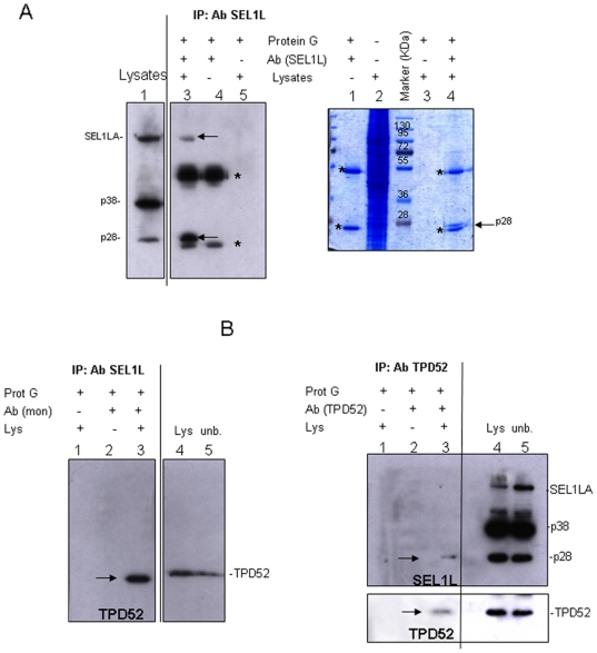
Figure 6. SEL1L and TPD52 immunoprecipitations assays.
A. SEL1LA and p28 immunoprecipitation analysis: Left panel: SKBr3 cell lysates (1.4 mg) were immunoprecipitated with monoclonal anti-SEL1L antibody (lane 3), resolved by SDS-PAGE (10%) and probed with monoclonal anti-SEL1L antibody. Lysate aliquots (50 µg, lane 1) were loaded to verify protein expression levels and immunoprecipitation efficiency. Arrows indicate the immunoprecipitated bands, asterisks (*) correspond to heavy and light chains. Absence of signal in controls (lanes 4 and 5) confirms the specificity of the immunoprecipitated bands. Right panel: SKBr3 cell lysates (7.0 mg) were immunoprecipitated with monoclonal anti-SEL1L antibody (lane 4), resolved by SDS-PAGE (10%) and stained with Coomassie brilliant blue. Asterisks (*) correspond to heavy and light chains. The arrow indicates the immunoprecipitated band analyzed by mass spectrometry. Absence of signal in controls (lanes 1 and 3) confirms the specificity of the immunoprecipitated band. B. Analysis of the interaction between SEL1L variants and TPD52: Left panel: SKBr3 cell lysates (1.4 mg) were immunoprecipitated with monoclonal anti-SEL1L antibody (lane 3), resolved by SDS-PAGE (10%) and probed with polyclonal anti-TPD52 antibody. Lysate aliquots (40 µg, lane 4) were loaded to verify protein expression levels and immunoprecipitation efficiency. Lane 5 corresponds to unbound aliquots of the samples loaded in lane 4 (40 µg). The arrow indicates the immunoprecipitated band. Absence of signal in controls (lanes 1 and 2) confirms immunoprecipitation specificity. Right panel: SKBr3 lysates (1.4 mg) were immunoprecipitated with polyclonal anti-TPD52 antibody (lane 3), resolved by SDS-PAGE (10%) and probed with monoclonal anti-SEL1L antibody. Lysate aliquots (40 µg, lane 4) were loaded to verify protein expression levels and immunoprecipitation efficiency. Lane 5 corresponds to unbound aliquots of the samples loaded in lane 3 (40 µg). The membrane was successively re-probed with anti-TPD52 antibody. Arrows indicate the immunoprecipitated bands. Absence of signal in controls (lanes 1 and 2) confirms immunoprecipitation specificity.

