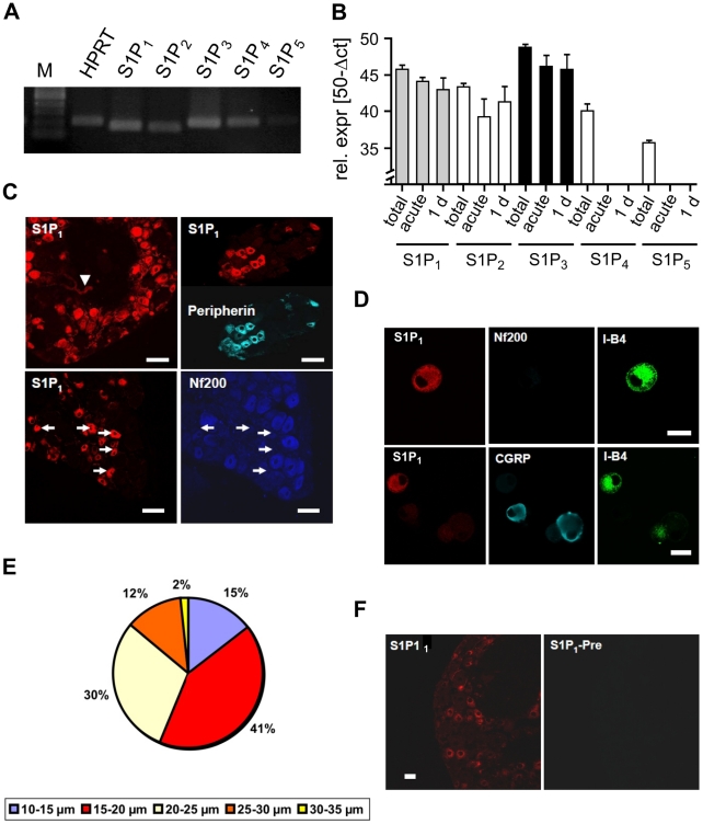
Figure 3. Expression of S1P1 receptors in sensory neurons.
(A) S1P receptor mRNA expression was detected with reverse transcription PCR in DRG explants. (B) Quantitative real-time PCR revealed expression of S1P1, S1P2 and S1P3 mRNA in DRG explants (total), acutely isolated neurons (acute) and 1-day-old cultures (1 d) (n = 5 experiments). In contrast, S1P4 and S1P5 mRNA levels were lower in DRG explants and absent in isolated neurons. (C) Immunoreactivity for S1P1 was present in neurons and intraganglionic capillaries (arrowhead). S1P1-IR was colocalized with immunoreactivity for peripherin, whereas S1P1-IR was absent in NF200-positive neurons. Scale bars = 50 µm. (D) S1P1 receptor colocalized with the small neuron marker I-B4 in the vast majority of cultured neurons but usually not with CGRP or Nf200, a marker for myelinated neurons (n = 4 experiments, scale bars = 20 µm). (E) Size distribution of S1P1-IR positive neurons revealed that S1P1-IR expressing cells are amongst the small diameter neurons (n = 6 experiments, 304 neurons). Only 2% of S1P1-IR+ neurons had diameters >20 µm. (F) Expression of S1P1 immunoreactivity was absent after preabsorption of the antibodies with the corresponding peptide.

