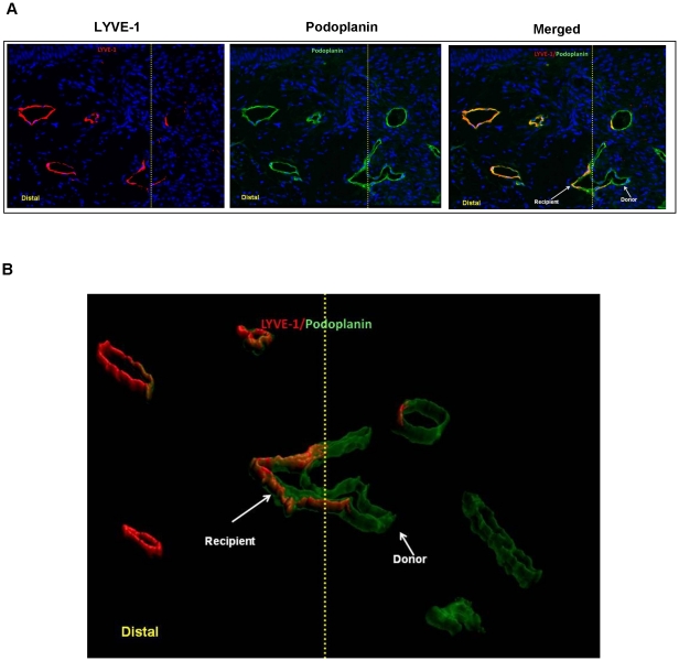
Figure 2. Lymphatic regeneration after tissue transfer occurs in part from spontaneous reconnection of recipient and donor lymphatic vessels.
A. Co-localization of LYVE-1 (left panel) and podoplanin (middle panel) of mouse tail sections harvested 6 weeks after surgery (40x). Overlay of images demonstrates connection of LYVE-1+ (i.e. recipient) and LYVE-1- (i.e. donor) derived podoplanin-stained lymphatics. Yellow line marks the junction of the skin graft and native tail skin. Distal junction of the skin graft and mouse tail is shown to the left. B. Three-dimensional rendering of podoplanin/LYVE-1 co-localization at the junction of the skin graft and native tail skin (yellow line). Note connection between LYVE-1+ and LYVE-1- lymphatic. Also note presence of both recipient (podoplanin+/LYVE-1+) and donor (podoplanin+/LYVE-1-) vessels in the section.

