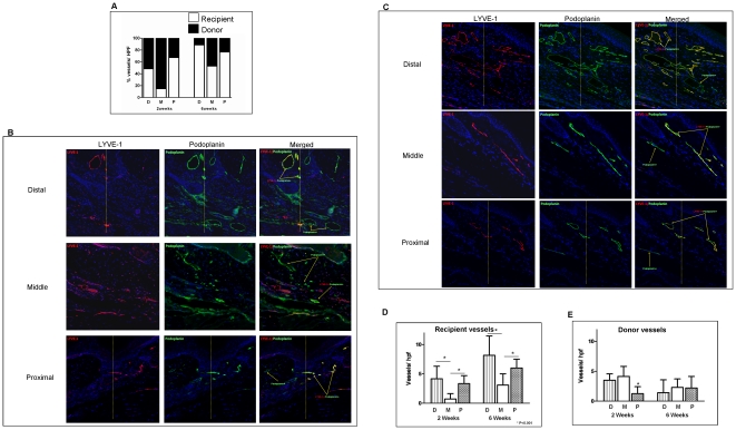
Figure 3. Both reconnection and infiltration of LYVE-1+ lymphatic vessels contribute to lymphatic regeneration after tissue transfer.
A. Bar graph depicting source of lymphatic vessels in various regions of the skin-grafted tissues 2 or 6 weeks after surgery. Origin of lymphatic vessels in various regions of the skin graft 2 or 6 weeks after surgery was determined using podoplanin/LYVE-1 co-localization in skin grafts harvested from LYVE-1 knockout mice and transferred to nude mice. Recipient lymphatic vessels were identified as podoplanin+/LYVE-1+ (white bars) while donor lymphatics were identified as podoplanin+/LYVE-1- (black bars). Recipient-derived lymphatic vessels were noted primarily in the distal and proximal portions of the graft at the 2-week time point and became more uniform in distribution by 6 weeks. Increased number of recipient vessels in the peripheral regions of the graft at the 6-week time point resulted in a relative decrease in the percentage of donor-derived lymphatics at this time. B, C. Representative images (10x) of LYVE-1 (red) and podoplanin (green) staining of the distal portion of the graft 2 (B) and 6 (C) weeks after surgery. Yellow line marks the junction of the skin graft (located to the right) and recipient (located to the left) tissues. D. Recipient lymphatic vessels (podoplanin+/LYVE-1+) in various regions of the skin graft 2 and 6 weeks after skin grafting. Note that the numbers of recipient vessels in the distal (D) and proximal (P) portions of the tail are significantly greater than the number of recipient lymphatics in the middle (M) portion of the graft at both the 2- and 6-week time points indicating ingrowth of vessels from the periphery (*p<0.05). In addition, the number of recipient lymphatics in various regions of the skin graft increased significantly when comparing 2- and 6-week time points indicating ingrowth of recipient vessels. E. Donor lymphatic vessels (podoplanin+/LYVE-1-) in various regions of the skin graft 2 and 6 weeks after skin grafting (*p<0.05). The number of donor vessels remained essentially unchanged at both time points indicating that donor lymphatics persist over time and do not proliferate after tissue transfer.

