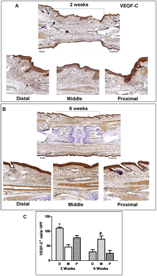
Figure 4. Lymphatic regeneration after tissue transfer is associated with expression of VEGF-C.
A. VEGF-C expression in skin-grafted tails 2 weeks after surgery. Representative low power (2x; upper panel) photomicrographs encompassing the skin-grafted area and distal/proximal portions of the recipient mouse-tail are shown. High power (20x) views of the distal and proximal junctions between recipient tissues and skin grafts are shown below. Black arrow shows large number of VEGF-C+ cells in the distal junction. Dashed box delineates skin-grafted area. B. VEGF-C expression in skin-grafted tails 6 weeks after surgery. Representative low power (2x; upper panel) and high power (20x) photomicrographs encompassing the skin-grafted area and distal/middle/proximal portions of the recipient mouse tails are shown. Dashed box delineates skin-grafted area. Note small amount of wound/skin graft contracture after repair. C. Cell counts per high power field of VEGF-C+ cells in the various tail regions (D = distal, M = middle, P = proximal) 2 and 6 weeks after surgery. Cell counts are means ± SD of at least 4 high power fields/mouse/time point. At least 6 mice were analyzed in each group (*p<0.05; #<0.01).

