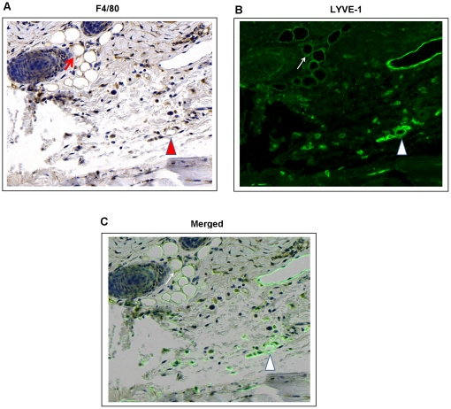
Figure 6. Recipient-derived macrophages express the lymphatic endothelial cell marker LYVE-1 after tissue transfer.
A, B, C. Representative photomicrograph (40x) demonstrating co-localization of F4/80 (A; brown immunostaining) and LYVE-1 (B; green fluorescent stain) in skin-grafted mouse tail sections 2 weeks after surgery. Arrows show co-localization of F4/80 and LYVE-1 in lymphatic capillaries. Also note scattered F4/80+/LYVE-1+ cells in the tail section. F4/80-/LYVE-1+ capillaries can also be appreciated (upper right corner).

