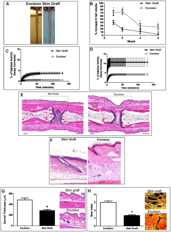
Figure 7. Spontaneous regeneration of lymphatics after tissue transfer can be used to bypass damaged lymphatics.
A. Gross photographs comparing nude mice that had undergone tail excision with (right) and without (left) skin grafting are shown 6 weeks after surgery. Note obvious difference in tail swelling. B. Tail volume measurements in nude mice that had undergone tail excision with or without skin grafting. Data are presented as percent change from baseline (i.e. preoperatively) with mean ± SD (*p<0.05). C, D. Representative lymphoscintigraphy of nude mice that had undergone tail excision with or without skin grafting. E. Representative photomicrograph (5x) of H&E stained tails sections from nude mice treated with (left) or without (right) 6 weeks after surgery. Dashed box delineates area of skin graft. Note decreased inflammation (cellularity) and dermal thickness in skin-grafted mice distal (to the left) of the wound. F. High power (40x) photomicrographs of tail skin harvested 5 mm distal to the excision site. Note decreased cellularity in skin-grafted section (left) as compared with excision section (right; arrow). Also note decreased dermal thickness. G. Dermal thickness measurements and representative figures (40x) in nude mice that had undergone tail excision with or without skin grafting 6 weeks following surgery (*p<0.05). H. Scar index measurements in tail tissues localized just distal to the site of lymphatic injury 6 weeks after treatment with excision with or without skin grafting. Representative Sirius red birefringence images are shown to the right. Orange-red is indicative of scar; yellow-green is consistent with normal (i.e. non-fibrosed) tissue (*p<0.01).

