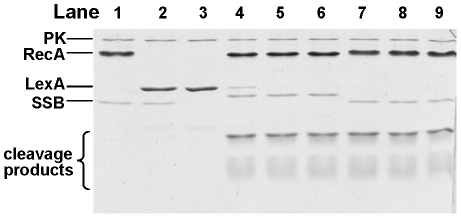
Figure 7. LexA cleavage promoted by RecANg and RecAEc.
LexA was incubated with RecA proteins in the presence of cssDNA, an ATP regeneration system [pyruvate kinase (PK) and phosphor(enol)pyruvic acid], and the cognate SSB protein over a 30 minute time course. Reactions were stopped and visualized on a 17% SDS-PAGE stained with Coomassie Brilliant Blue. Lanes 1–3 are negative controls (incubated for 30 min) which lack various protein components of the reaction and are as follows: 1) lacks only LexA; 2) lacks RecA; 3) lacks RecA and SSB; lanes 4–6 contain the complete reaction and RecANg and SSBNg proteins, with time points taken at 5, 15, and 30 min; lanes 7–9 contain the complete reaction and RecAEc and SSBEc proteins with time points taken at 5, 15, and 30 min. The two LexA cleavage products are visible at the bottom of the gel. The faint band in lane 3 that migrates slightly slower than the LexA cleavage products is likely a breakdown product of LexA.

