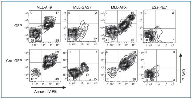
Figure 4.
Increased Annexin V labeling in Dot1l deleted cells immortalized by MLL-AF9, MLL-GAS7, or MLL-AFX but not by E2a-Pbx1.
Immortalized hematopoietic cells expressing the indicated oncogenes were transduced with GFP or Cre-GFP, labeled with Annexin V-PE/7-AAD, and analyzed by flow cytometry 5 days after transduction. The Annexin V-positive/7-AAD-negative cells in the lower right quadrant and the Annexin V–positive/7-AAD-positive cells in the upper right quadrant represent early apoptotic and late apoptotic/necrotic cells, respectively. GFP+ cells are presented as dual parameter contour plots and the percentage of cells in each quadrant is indicated.

