Fig. 1.

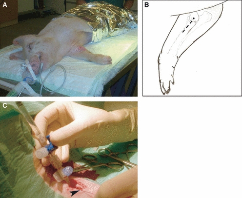
The inoculation procedure. (A) Anaesthetized pig in right lateral recumbency with the right medial ante-brachium prepared for surgery. (B) Schematic drawing, the skin incision is indicated by the punctured line and the asterisk (*) shows the location of the bone protuberance epicondylus medialis humeri. (C) Inoculation through a catheter equipped with a three-way stopcock inserted into the right brachial artery fixated with a ligature ( ).
).
