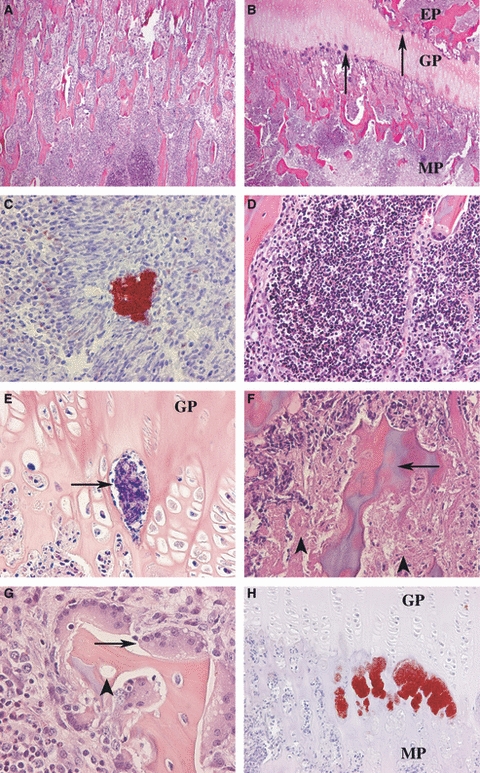
Fig. 2.

Histopathology in pigs inoculated intra-arterially (a. brachialis dextra) with 50 000 CFU/kg BW. (A) Radius: a microabscess located deep in the metaphyseal area, H&E. (B) Metacarpale III: colonies of bacteria (→) are present at the junction between the growth plate (GP) and the metaphysis (MP) and the epiphysis (EP), H&E. (C) Epiphysis of phalanx proximalis III: multiple Staphylococcus aureus bacteria are seen centrally in the microabscess. Surrounding cells are arranged in a pattern of palisades. Immunostaining for S. aureus. (D) The centre of the microabscesses was made up by accumulation of neutrophils, H&E. (E) Within the blood vessels of the growth plate (GP), fibrin deposition was sometimes observed (→), phosphotungstic acid haematoxylin (PTAH). (F) Osteonecrosis (→) was often present just beneath the growth plate together with areas of necrotic bone marrow cells ( ), H&E. (G) Trabeculae with empty lacunae (
 ) were typically surrounded by bone resorbing osteoclasts (→), H&E. (H) Multiple S. aureus bacteria were often identified in connection with the capillary loops at the junction between the growth plate (GP) and the metaphysis (MP). Immunostaining for S. aureus.
