Fig. 1.

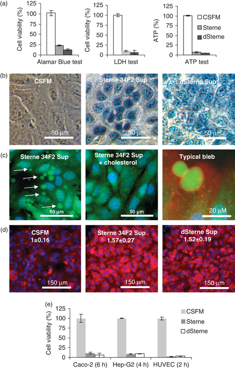
Bacillus anthracis generates toxic secreted products in static cultures. (a, b) Bacteria were grown in the CSFM. The viability of HSAECs relative to untreated controls was tested after incubation with Sups for 2 h at 37°C, 5% CO2 using the Alamar Blue test (a, left panel), LDH test (a, middle panel), intracellular ATP test (a, right panel), and the Trypan Blue permeability test (b). (c) Formation of membrane blebs in HSAECs incubated with Sterne 34F2 Sup. HSAECs were loaded with Calcein AM (5 μM, green), Hoechst 33342 (2.5 μg mL−1, blue), and wheat germ agglutinin conjugated with Alexa Fluor 555 (5 μg mL−1, red), washed with HBSS, and treated with the Sterne Sup for 30 min. The cells demonstrate a transient (up to 30-min postexposure) appearance of numerous membrane blebs (left panel, arrows). The addition of CD-cholesterol (1 μg mL−1) to the Sup for 30 min completely prevents blebbing (middle panel). A typical bleb (right panel) contains a part of the cell cytoplasm (green) surrounded by the cytoplasmic membrane (red). Similar results were obtained with dSterne Sup (not shown). (d) HSAECs grown on slides were incubated with Sups for 2 h at 37°C, 5% CO2, stained with Annexin V-conjugated Cyt3.18 dye (red) and nuclear DAPI dye (blue) for 10 min, fixed, and mounted for imaging. Numbers show the mean intensity of fluorescence (± 95% confidence interval) inred channel relative to the control cells incubated in CSFM. The intensity was evaluated with nis elements software (Nikon) using 10 independent fields of view in three different culture wells for each experimental condition. (e) Viability of Caco-2, HEP-G2, and HUVEC cells in the Alamar Blue test relative to the untreated control cell after the indicated times of exposure to Sups at 37°C, 5% CO2. Error bars represent 95% confidence interval of mean.
