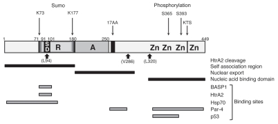
Figure 2.
Functional motifs and interactions of WT1. A linear schematic of WT1 is shown with numbering indicating amino acids. Zn is zinc finger, A is the activation domain, R is the repression domain, SD is the suppression domain. The alternative splice sites (17AA and KTS) are indicated. The HtrA2 cleavage sites, the self-association, nuclear export and nucleic acid-binding domains are indicated below and post-translational modifications (sumoylation and phosphorylation) are shown above. The binding sites in WT1 for BASP1, HtrA2, Hsp70, Par-4 and p53 are indicated.

