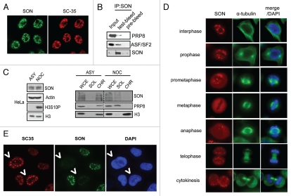
Figure 1.
SON is a component of spliceosomes. (A) SON colocalises with spliceosome marker SC-35. HeLa cells grown on coverslips were fixed, permeabilised and immunostained with indicated antibodes. (B) SON physically interacts with spliceosome components. Nuclease-treated HeLa cell extract was subjected to co-immunoprecipitation using Protein A agarose beads conjugated with anti-SON antibodies or pre-bleed control. Immunoprecipitates were washed with NETN buffer twice, eluted by boiling and separated by SDS-PAGE. Western blotting was performed using indicated antibodies. (C) SON associates with chromatin during interphase and is released during mitosis. HeLa cells were either untreated (ASY) or arrested in mitosis (NOC) by incubating with 100 ng/ml nocodazole for 16 hr. Cell lysates were biochemically fractionated according to Methods. Western blot analysis of SON expression in whole cell extract (WCE), soluble (SOL) and chromatin-enriched fractions (CHR). (D) SON concentrates in nuclear speckles and disperses during metaphase. Indirect immunofluorescence staining showing SON localization at different cell cycle phases. (E) SON deficiency compromises spliceosome function. HeLa cells treated with control or SON-specific siRNAs were mixed, seeded onto coverslip and processed 24 hr post-transfection. Indirect immunostaining was performed as described in Methods using indicated antibodies. SON-depleted cells are marked by arrows.

