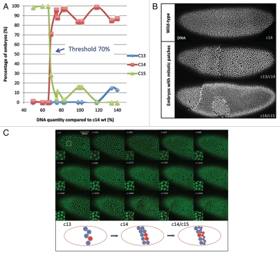
Figure 1.
Patchy cell cycle pause during Drosophila mid-blastula transition. (A) Cell cycle behaviors of embryos with different amount of DNA compared with wild-type. Below the threshold of 70%, embryos undergo one extra division. Above the threshold, embryos pause at cycle 14 as wild-type. (B) Embryos with threshold DNA form mitotic patches during MBT. Embryos are fixed and stained with Hoechst. (C) Snapshot of a movie of mitotic patch formation. The white square region is enlarged for better visualization. Red dots label the lineage of a nucleus. The cartoon below depicts the lineage history of a nucleus colored in red at the patch boundary. Scale bar = 50 µm.

