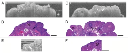
Figure 2.
Acyclic ovary. (A) OCT image of acyclic VCD/Con ovary and (B) corresponding histology. (C) OCT image of cyclic Con/DMBA ovary and (D) corresponding histology. (E) Magnification of Figure 2A acyclic VCD/Con ovary and (F) corresponding magnified histology. S, stroma; DF, degenerating follicles; SF, secondary follicle; F, infiltrating fat; CL, corpus luteum with cyst; arrow, epithelial invagination; asterisk, vascular spaces. OCT image and histology are to scale. Scale bar: 500 µm.

