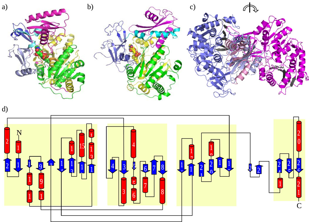
Figure 3.
Structure of FAAL from E. coli and L. pneumophila. (a) EcFAAL monomer showing location of acyl adenylate (sphere model) (b) Structure of LpFAAL monomer showing location of acyl adenylate (sphere model). N-terminal domain has three subdomains 1, 2, 3 colored in green, yellow, and pale violet, respectively. Insertion motif, hinge region, and C-terminal domain are colored in dark blue, light cyan and magenta, respectively. (c) EcFAAL (blue) and LpFAAL (magenta) overlaid at C-terminal domain. Sheet A of N-terminal domain is colored in black and is rotated about 180 degrees about the vertical axis. (d) Topology diagram of EcFAAL. N-terminal subdomain and C-terminal domain are highlighted in light yellow.

