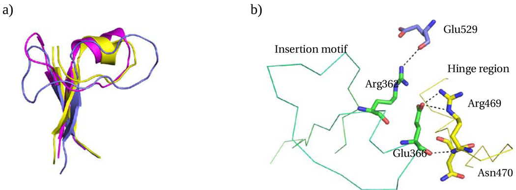
Figure 5.
Insertion motif. a) Insertion motifs of EcFAAL (magenta), LpFAAL (blue) and MtFAAL (yellow) are superposed. b) The C-alpha trace of EcFAAL insertion motif (green) and hinge region (yellow) is shown with the critical interactions between insertion motif and the C-terminal domain or hinge region highlighted.

