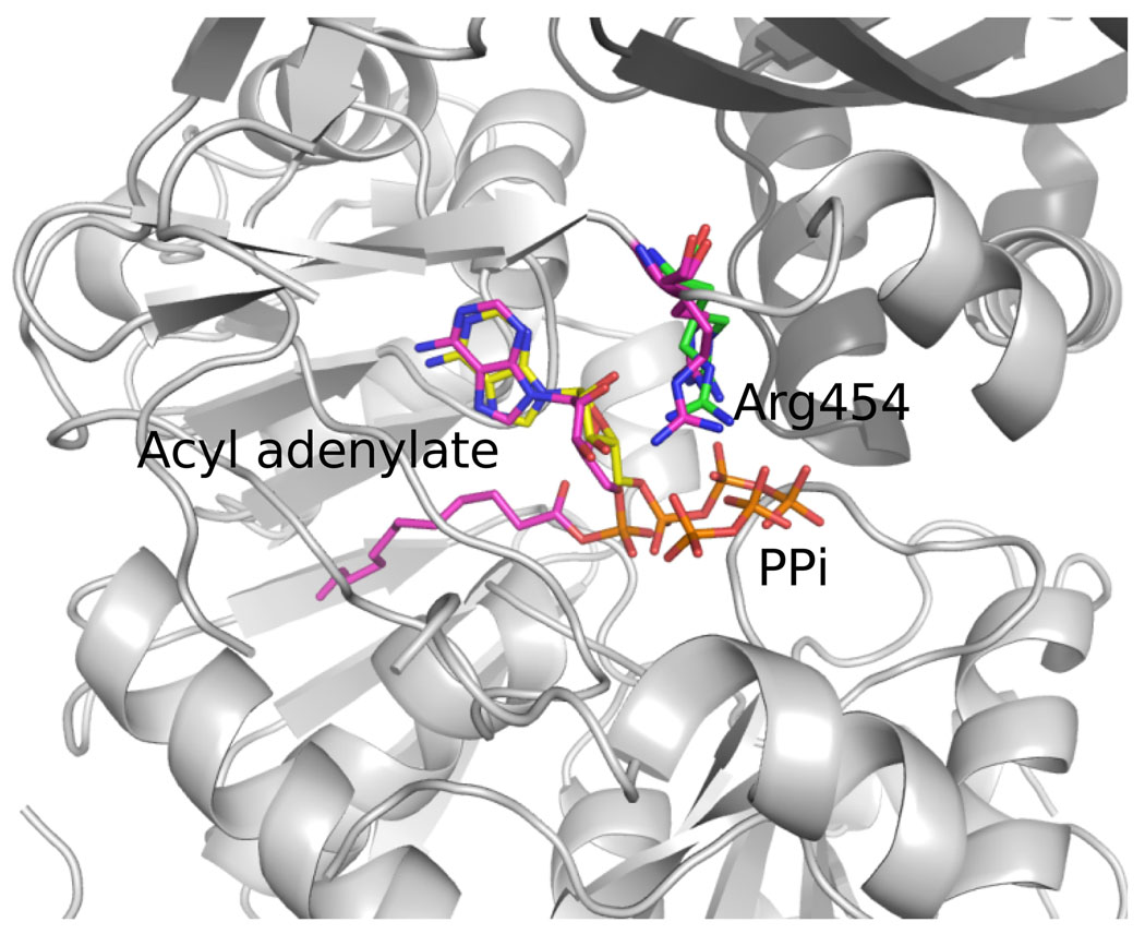
Figure 7.
Overlay of LpFAAL with acyl adenylate/PPi bound and HsFACL with ATP bound. The N- (grey) and C-(darker grey) terminal domain of LpFAAL are shown as cartoons. Acyl adenlylate, PPi and Arg454 are colored in magenta. HsFACL were only presented with ATP and Arg461, colored in yellow and green, respectively.

