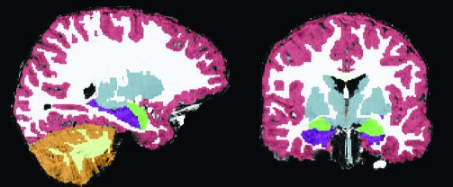
Figure 1.
Example segmented brain. On the left is a sagittal view and on the right is a coronal view demonstrating tissue segmentation of a single participant’s brain. Segmentation includes cortical gray (red) and white matter (white) segmentation, and subcortical structures, the majority of which are colored in gray. Subcortical regions of interest are highlighted in bright colors (the hippocampus colored in purple and the amygdala in green).

