Figure 4.

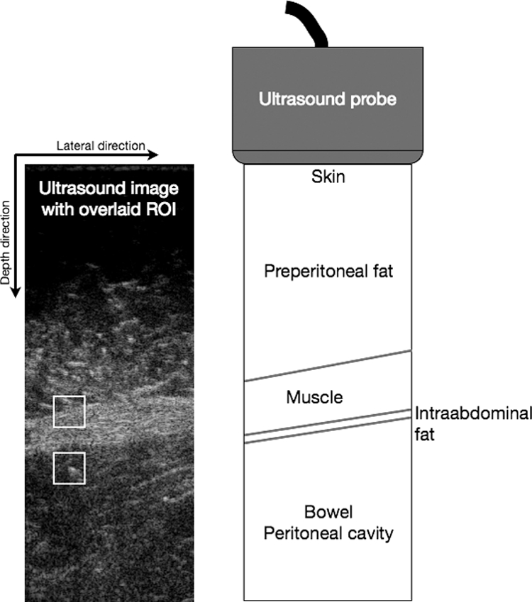
The ultrasound image contains overlays of the defined ROIs, the top one set in the abdominal wall and the lower ROI set in the underlying bowel. The drawing indicates the various structures covered by the image.

The ultrasound image contains overlays of the defined ROIs, the top one set in the abdominal wall and the lower ROI set in the underlying bowel. The drawing indicates the various structures covered by the image.