
Fig. 2.
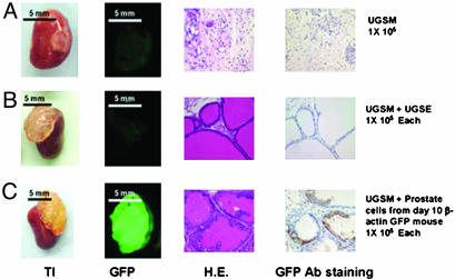
Dissociated prostate epithelial cells combined with UGSM can regenerate the prostatic-like tissue. UGSE (1 × 10⁵) or 1 × 10⁵ 10-d-old β-actin GFP mouse prostate epithelial cells were mixed with 1 × 10⁵ UGSM, and the regeneration protocol was carried out as described in Materials and Methods and Fig. 1. Mice were killed 3 mo after the surgery, and regenerated tissues from the kidney capsules were collected. (A) UGSM (1 × 10⁵) alone was used as a negative control. (B) Graft initiated with 1 × 10⁵ UGSM and 1 × 10⁵ UGSE. (C) Graft composed of 1 × 10⁵ UGSM and 1 × 10⁵ dissociated 10-d-old β-actin GFP prostate epithelial cells. (A and B) From left to right: transilluminated image (TI) and green fluorescent image (GFP) of regenerated grafts; hematoxylin/eosin staining (H.E.), and the GFP antibody staining (GFP Ab staining) of regenerated tissue sections. GFP protein was detected by immunohistochemistry with rabbit polyclonal antibody against GFP (Abcam, 1:300 dilution) with the Envision+ system (DAKO). Photos were taken with a Leica MZFLIII dissecting microscope (×10). (Bars = 5 mm.)