
Fig. 4.
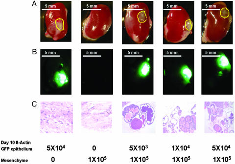
Number of postnatal day 10 epithelial cells required for effective regeneration. UGSM cells (1 × 10⁵) were mixed with 10-d-old β-actin GFP mouse prostate epithelial cells ranging from 5 × 10³ to 5 × 10⁴, and the regeneration protocol was carried out as described in Materials and Methods and Fig. 1. Mice were killed 1 mo after surgery, and regenerated tissues from the kidney capsules were collected. UGSM alone and β-actin GFP mouse prostate epithelial cells alone were used as controls. (A) Transillumination. (B) Green fluorescence image of the regenerated tissues under kidney capsules. (C) Hematoxylin/eosin staining of the regenerated tissue sections. Numbers below the panels indicate the cell number of the UGSM and the prostate epithelial cells. Yellow circles in A show the positions of the regenerated tissues. A and B photos were taken with a Leica MZFLIII dissecting microscope (×10). (Bars = 5 mm.) (C) Photo was taken with a Nikon Diaphot phase contrast microscope (×100).