Fig. 4.
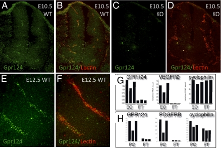
Gpr124 is expressed in normal developing CNS vessels of E10.5 and E12.5 embryos. Fluorescent RNA in situ hybridization with a probe for Gpr124 (green) coupled with fluorescent histochemical staining of endothelial cells by GS lectin I (red) in cross-sections through the neural tube of E10.5 WT embryos (A and B) showed Gpr124 expression within the developing CNS vasculature. Gpr124 expression was not detected in Gp124 KO embryos (C and D). Gpr124 (green) continued to be expressed within the vasculature of the CNS in WT E12.5 embryos (E and F). RT-PCR measurements on platelet endothelial cell adhesion molecule–positive endothelial cells and PDGF receptor β–positive pericytes isolated by FACS from the brains of WT embryos at E15.5 demonstrated that both endothelial cells (EC) and pericytes (PC) have enriched expression of Gpr124 relative to nonendothelial cells [“flow through” (FT)] and nonpericyte cells (FT), respectively (G and H). Control probes for VEGF receptor 2, PDGF receptor β, and cyclophilin confirmed the endothelial identity of isolated cells, the pericyte identity of isolated cells, and the integrity of mRNA samples, respectively. The y axis scale is proportional to RNA copy number.

