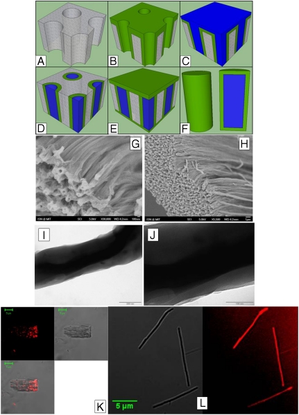
Fig. 1.
Schematic of the microworm fabrication process. The bare AAO template (A) is first conformally coated with the iCVD hydrogel layer (B). The optode solution is filled in the pores of the template (C) and the excess optode and the hydrogel layer is etched away (D). A final hydrogel layer is deposited on both sides of the template to cap the optode (E). As the final step, the template is dipped in the HCl solution to etch the AAO template and release the microworms (F). (G and H) SEM images of the microworms in the dehydrated state. (I and J) TEM images of the microworms. The inner optode core and the outer hydrogel layers are visible. The hydrogel coating has a thickness of 50 ± 10 nm. (K) Confocal images of control sensors without hydrogel coating. Large piece of template, that is not completely etched, containing spots of hydrophobic optode is shown. Fluorescence (Upper Left), bright field (Upper Right), and overlay (Lower Left) are shown. (L) Confocal image of microworms. Shown are the brightfield (Left) and fluorescent (Right) images of microworms with scale bar.

