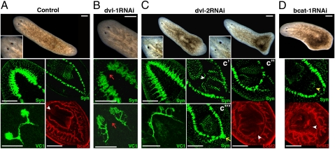
Fig. 4.
Smed-dvl paralogs are functionally specialized. (A) Stereomicroscopic image of a control animal (magnified view of the eye region shown at Bottom Left), with anti-Syn staining of the anterior and posterior regions, anti-VC1 staining of the visual system, and anti-bcat2 staining showing the two posterior branches and the pharynx. The white arrowhead indicates the anchoring of the pharynx. (B) dvl-1 RNAi animal lacking the ATC and with aberrant visual system regeneration (red arrows). (C) Analysis of a dvl-2 RNAi animal. Note the poor and asymmetrical differentiation of the eyes. An ectopic posterior brain is indicated with a yellow arrow, and the anchoring of the ectopic pharynges is marked with white arrowheads. (D) Low-dose bcat-1 RNAi animal showing the differentiation of an ectopic mouth (yellow arrowhead) and an ectopic pharynx primordium (white arrowhead). All images correspond to regenerating trunk pieces. (Left) Anterior. Anti-Syn, bcat2, and anti-VC1 staining images correspond to confocal z-projections. bcat1, Smed–β-catenin1; bcat2, Smed–β-catenin2; dvl-1, Smed-dvl-1; dvl-2, Smed-dvl-2; Syn, synapsin; VC1, arrestin. (Scale bar: 250 μm.)

