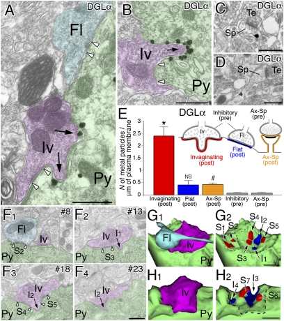
Fig. 2.
Silver-enhanced immunogold electron microscopy demonstrating intensive accumulation of DGLα at invaginating synapses on BA pyramidal neurons. (A and B) DGLα is concentrated on the somatic membrane of pyramidal neurons (Py) that apposes to invaginating terminals (arrows, Iv). However, no particular labeling is seen for terminals forming the flat type of perisomatic synapses (Fl). Open arrowheads indicate the edges of symmetrical synaptic contact. (C and D) A few metal particles are detected in the head and neck portions of dendritic spines (Sp) forming asymmetrical synapses with putative excitatory terminals (Te). (E) Labeling density of metal particles for DGLα in the postsynaptic membrane of invaginating, flat, or axospinous synapses (colored portions for quantitative measurement), and in the presynaptic membrane of inhibitory (symmetrical) or axospinous synapses (gray portions). Using 50 electron micrographs taken from three mice, we analyzed the total number of 19, 18, 200, 35, and 189 synaptic elements. *P < 0.01 compared with all other labelings; NS, P > 0.05 compared with presynaptic labeling at inhibitory synapses; #P < 0.01 compared with presynaptic labeling at axospinous synapses, Mann–Whitney U test. (F–H) Serial electron microscopic images (F) and 3D reconstruction of invaginating perisomatic synapses (G and H) in BA pyramidal neurons. G was reconstructed from 24 serial sections, including those shown in F. Invaginating terminal (Iv), flat terminal (Fl), and pyramidal neuron soma (Py) are pseudocolored in purple, light blue, or green, respectively. Individual sites of invagination (I1–I4) and synaptic contact (S1–S7) are indicated by arrows or open arrowheads, respectively, in F, and by blue or red area, respectively, in G and H. (Scale bars: 500 nm.)

