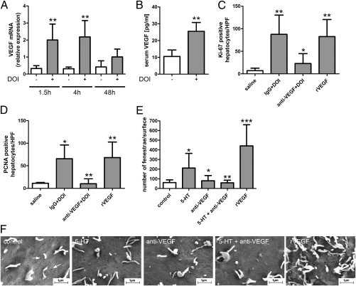
Fig. 4.
VEGF mediates DOI-induced opening of fenestrae and liver regeneration. (A) Gene expression of VEGF in old animals treated with (gray bars) and without (white bars) DOI (n = 5). **P < 0.008 vs. animals without DOI. (B) Serum VEGF levels in old mice after treatment with and without DOI for 48 h (n = 4). **P = 0.007. (C and D) Assessment of liver regeneration 48 h after partial hepatectomy in old mice in the presence of unspecific IgG or anti-VEGF antibodies followed by DOI injection. Administration of recombinant VEGF alone followed by partial hepatectomy also enhanced liver regeneration. (C) Quantification of Ki-67 immunostainings (n = 5). **P < 0.001 vs. control; *P < 0.05 vs. IgG + DOI. (D) Quantification of PCNA immunostainings (n = 5). **P < 0.001 anti-VEGF + DOI vs. IgG + DOI and recombinant VEGF vs. control; *P < 0.05 vs. control. (E and F) Quantification and photographs of fenestrae (white arrows) in the endothelial cell line SK HEP-1 incubated with 5-HT, anti-VEGF antibodies alone, anti-VEGF antibodies plus 5-HT, or recombinant VEGF. *P < 0.05 vs. control or 5-HT; **P < 0.001 vs. 5-HT; ***P < 0.001 vs. control.

