Fig. 2.
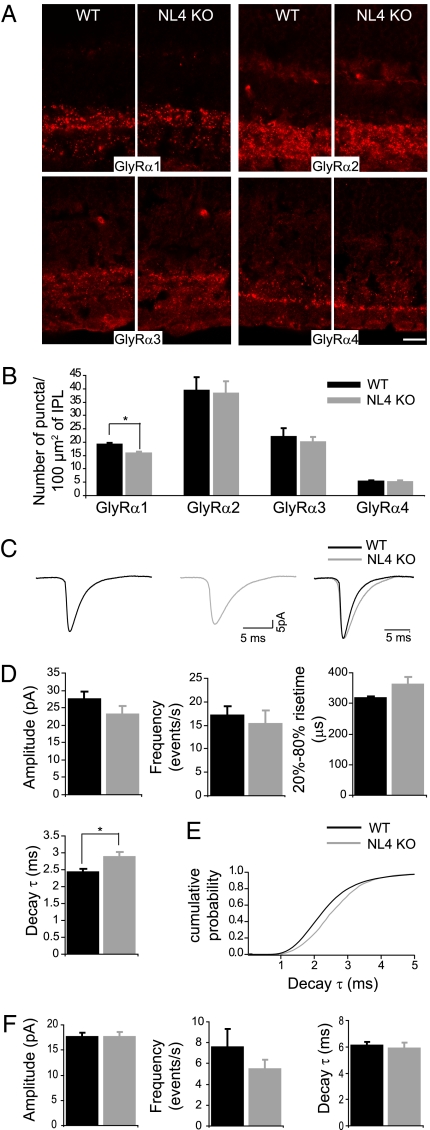
NL4 loss causes alterations of the glycinergic circuit. Distinct populations of GlyRs bearing α1–α4 subunits were similarly distributed in WT and NL4-KO retinae (A). Quantitative analysis uncovered a specific reduction in the number of GlyRα1 clusters in NL4-KO retinae (B). Glycinergic (C–E) and GABAergic (F) mIPSCs were recorded from WT and NL4-KO RGCs. (C) Example traces of WT and NL4-KO glycinergic mIPSCs. (D) Glycinergic mIPSCs from NL4-KO RGCs were not significantly different in frequency, amplitude, and rise time, but showed significantly slower decay kinetics compared with WT. Cumulative histogram of decay time constant (τ) values of glycinergic mIPSCs (E) showed a rightward shift in the KO. Amplitude, frequency, and decay time constant (τ) of GABAergic mIPSCs were similar in WT and NL4-KO RGCs. (Scale bar: 5 μm.)

