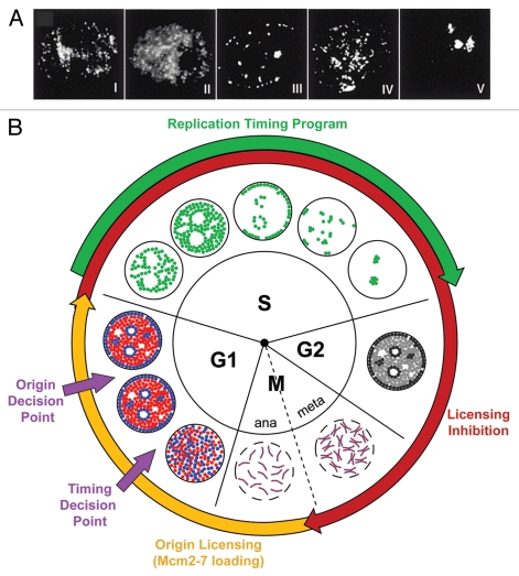
Figure 1.
The replication timing program. (A) CHO cells, previously synchronized at the G1/S border and released into S-phase for various times, were pulse labelled with BrdU for 5 min and stained with anti-BrdU antibodies to visualize patterns of DNA replication. Shown are characteristic examples of each of the 5 labeling patterns (Types I–V). Reproduced from Dimitrova & Gilbert 1999. (B) The replication cycle—the establishment and execution of replication timing in the cell cycle. The innermost wheel marks the phases of the cell cycle and the outermost wheel the key stages of DNA replication, where the periods of licensing competence and inhibition are shown in yellow and red respectively and the execution of the replication timing program during S-phase is shown in green. Key nuclear transitions are represented diagrammatically in the central portion. During mitosis, paired sister chromatids aligned on the metaphase plate are separated to opposite poles of the dividing cells upon entry into anaphase. Late mitotic chromosome decondensation then readies DNA for emergence into G1. In early G1, after nuclear envelope reformation (continuous black line), regions of euchromatin, shown in red and heterochromatin, shown in blue, initially randomly dispersed, move to occupy specific domains within the nucleus. During S-phase, replication foci, shown in green, are activated in accordance with the execution of the timing program. Post-replicative nuclei pass through G2 into mitosis, where chromosome condensation and nuclear envelope breakdown (dashed black line) ready the cell for sister chromatid separation and cell division. The execution of the timing decision point and the origin decision point are marked.

