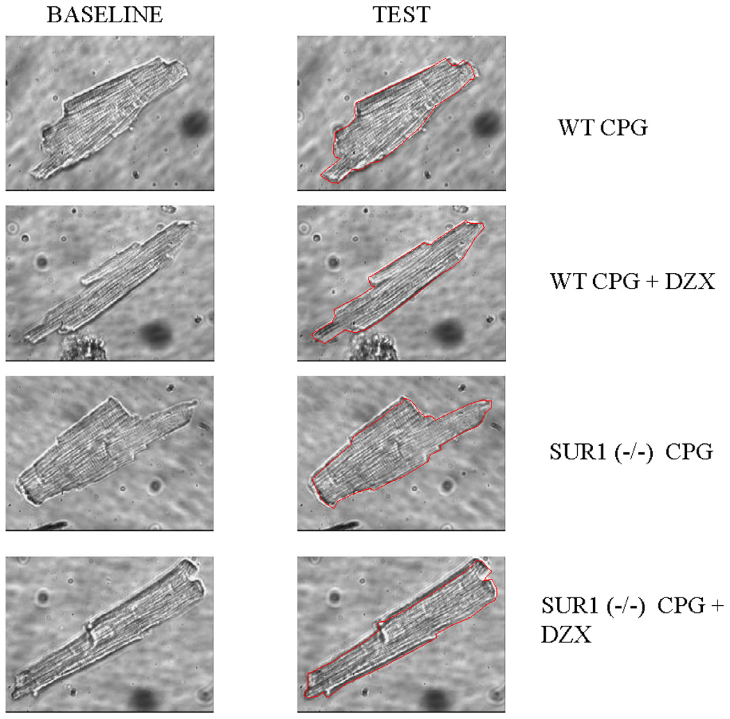
Figure 5.

Representative isolated wild type or SUR1 (−/−) myocytes at baseline volume and following exposure to test solution via inverted microscopy. Myocytes were initially exposed to normal Tyrode’s solution for 5 minutes for baseline volume measurement. Myocytes were then exposed to test solution (hyperkalemic cardioplegia or hyperkalemic cardioplegia + 100µM diazoxide). The red line drawn on the representative test cell represents the outline of that cell’s baseline volume. Significant volume increase was noted in response to cardioplegia in the wild type (WT) and SUR (−/−) cells, and this was eliminated with the addition of diazoxide in the WT cells only. WT is wild type, CPG is cardioplegia, and DZX is diazoxide.
