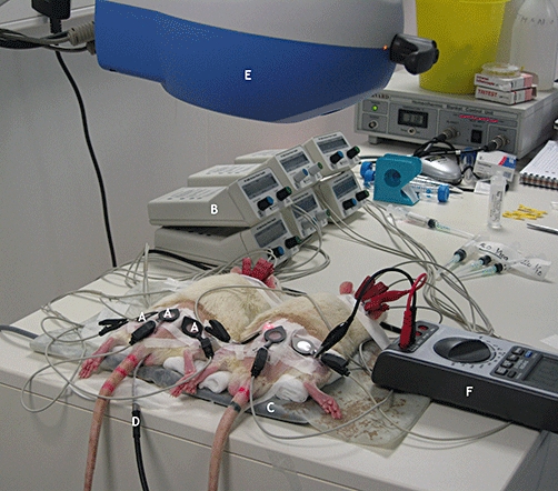
Figure 1.

Experimental setup: three iontophoresis chambers (A) connected to power supplies (B) were placed on the back and the hind legs of two shaved rats placed in the prone position on a thermal pad (C), temperature being maintained at 37.5°C and adjusted using a rectal probe (D) in one rat. Laser Doppler imaging (E) was performed over the area including all iontophoresis chambers. Cutaneous resistance was determined during iontophoresis with an amperometric biosensor unit (F).
