
Figure 2.
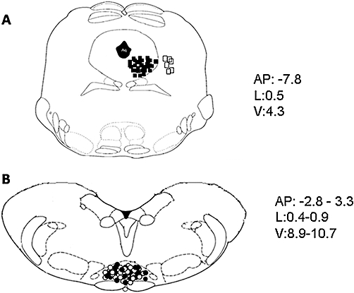
Schematic illustration of the location of periaqueductal gray (PAG) microinjection sites (A) and rostral ventromedial medulla (RVM) ON or OFF cell recording sites (B). Vehicle or drug microinjections were performed in the ventrolateral (vl)-PAG (filled squares) (A). Open squares indicate the microinjection sites performed outside the vl-PAG, which were neither associated with change in RVM cell activity nor with tail-flick latency. Moreover, cell recordings performed by lowering a tungsten electrode into the RVM and ON cells (filled circles) or OFF cell (open circles) sites (B) are shown. Many sites are not shown to avoid symbol overlapping. Distances (mm) from the interaural line are indicated.