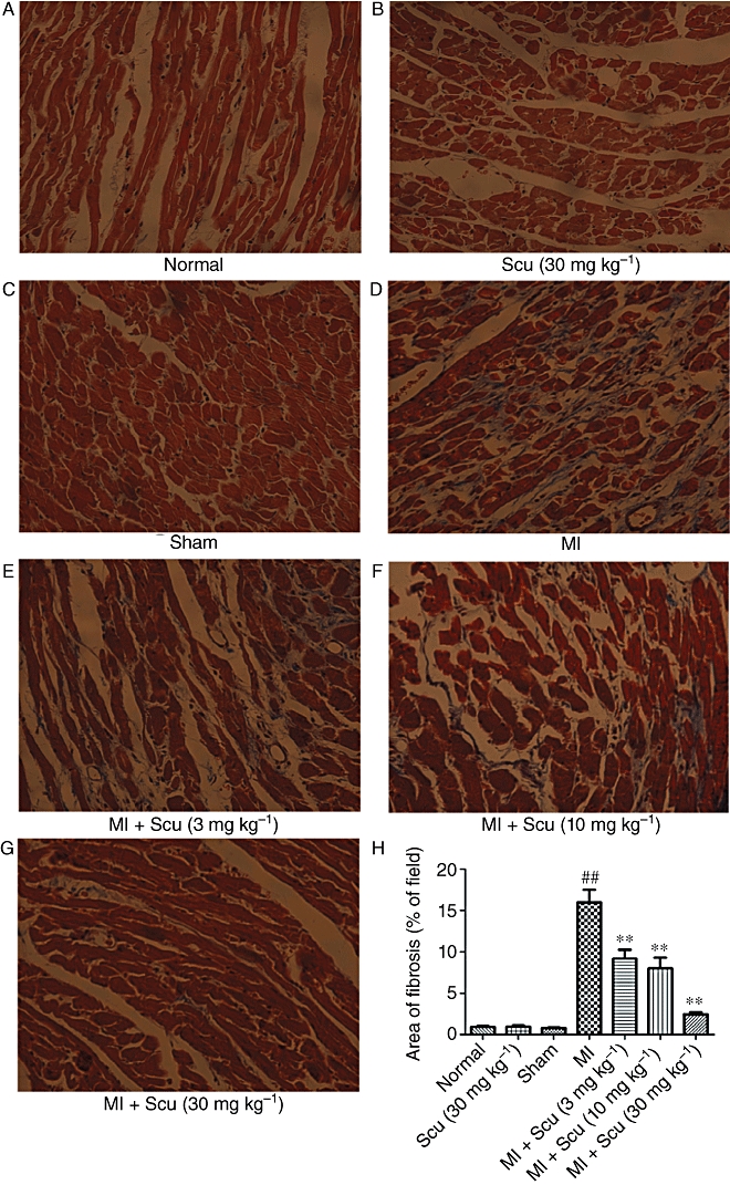
Figure 4.

Effects of scutellarin (Scu) on the deposition of collagen in the peri-infarct region during myocardial infarction (MI) in rats. (A–G) Representative sections of heart stained with Masson's trichrome viewed at a magnification of 200×. The fibrotic area is stained blue and the viable area red. (H) Collagen deposition was quantified by automated image analysis and expressed as percentage of tissue area. Data are expressed as mean ± SEM, n = 5. ##P < 0.01 versus sham; **P < 0.01 versus MI.
