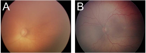
Figure 2.
Examples of disagreements. In (A), ROP was documented by image-based exam (arrow point at vertical white line denoting peripheral disease) but not by ophthalmoscopy. In (B), there was disagreement regarding whether dilation and tortuosity of vessels was sufficient to meet criteria for severe ROP.

