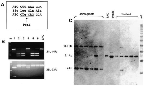
Figure 3.
(A) The box shows the exon sequence around the introduced nucleotide change and its translation into protein. The first line shows the original sequence. The nucleotide change, in the third line, is shown in lower case letters and the new PstI site is underlined. (B) PCR amplification of BAC-shuttle co-integrants using primers shown on the right (see Fig. 2) and digested with PstI. Clones 2, 3, 5 and 6 contained the mutation on the ‘left’ arm of the co-integrant and clones 1 and 4 on the ‘right’ arm. (C) Southern blot of the co-integrant clones, the original BAC, the shuttle vector and resolved BAC clones carrying the mutation, were digested with EcoRI and hybridised with exon 7.

